Current research topics:
| |
|
| |
|
|
Biomineralization in magnetotactic bacteria |
| |
|
|
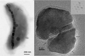
Magnetotactic bacteria provide probably the best studied example of
biomineralization: they form intracellular magnetosomes which are
nanocrystals of magnetic minerals (magnetite or greigite) surrounded by
a biological membrane. The grains of magnetic minerals are typically 35
to 120 nm in diameter, which makes them single-domain (SD)
ferrimagnets. |
| |
|
|
We study the minerals produced by magnetotactic bacteria in order to
gain insight into the biomineralization process and to determine the
special phyiscal and chemical properties of the magnetic nanocrystals.
We collaborate with microbiologist colleagues who isolate and culture
various strains of bacteria and create genetically modified mutants.
These samples allow us to assess the changes in biomineral properties
as a result of mutations in the genes that are responsible for
controlling the biomineralization process through magnetosome membrane
proteins. We use various transmission electron microscopy techniques to
study the sizes, morphologies, structures, compositions, and magnetic
properties of the particles. |
 |
| |
|
|
Currently involved in this research at the UP: Éva Bereczk-Tompa, PhD student. |
| |
|
|
Collaborations: Prof.
Dirk Schüler, University of Bayreuth (genetically modified mutants of
Magnetospirillum gryphiswaldense); Prof. Dennis Bazylinski, University
of Nevada at Las Vegas, and Dr. Christopher Lefèvre, CNRS Cadarache,
France (various types of isolated and cultured magnetotactic bacteria);
Prof. Richard Frankel, California Polytechnic State University, San
Luis Obispo (physics of magnetotaxis). |
| |
|
|
Recent papers:
|
|
Lohße, A., Borg, S., Raschdorf, O., Kolinko I.,Tompa, É., Pósfai, M., Faivre, D., Baumgartner, J. and Schüler, D. (2014) Genetic
dissection of the mamAB and mms6 operons reveals a gene set essential
for magnetosome biogenesis in Magnetospirillum gryphiswaldense. Journal of Bacteriology, JB.01716, doi: 10.1128/JB.01716-14. |
|
Kolinko, I., Lohße, A., Borg, S., Raschdorf, O., Jogler, C., Tu, Q.,
Pósfai, M., Tompa, É., Plitzko, J.M., Brachmann, A., Wanner, G.,
Müller, R., Zhang, Y. and Schüler, D. (2014) Biosynthesis of magnetic nanostructures in a foreign organism by transfer of bacterial magnetosome gene clusters. Nature Nanotechnology,
9, 193-197. doi: 10.1038/nnano.2014.13. |
|
Pósfai, M., Lefèvre, C.T., Trubitsyn, D., Bazylinski D.A., and Frankel, R.B. (2013) Phylogenetic significance of composition and crystal morphology of magnetosome minerals. Frontiers in Microbiology, 4, 344. doi: 10.3389/fmicb.2013.00344. |
| |
|
| |
|
|
Characterization of magnetic nanoparticles |
| |
|
|
Magnetic nanoparticles are interesting for science and technology for a
number of reasons. Our main interest is in biomineralization (see
above), but technological and medical applications are also extremely
important. For such applications the nanoparticles need to be
characterized as best as is possible, and no other technique can match
the versatility of the transmission electron microscope (TEM). | |
| |
|
|
The study of the structure and chemistry of nanometer-sized particles
poses serious challenges for the electron microscopist. We use some
established TEM techniques such as amplitude-contrast and
high-resolution imaging and selected-area electron diffraction for the
analysis of particle sizes and structures. Compositions are analyzed
using energy-dispersive spectrometry and electron energy-loss
spectroscopy and imaging in the TEM. However, the magnetosomes are
embedded in cells, they are small enough (<10 nm, especially in some
genetically modified mutants) to cause significant fringe
delocalization in HRTEM images. Obtaining meaningful elemental
compositions is also difficult. This type of work thus needs experience
with the TEM and patience with carrying out the measurements. In
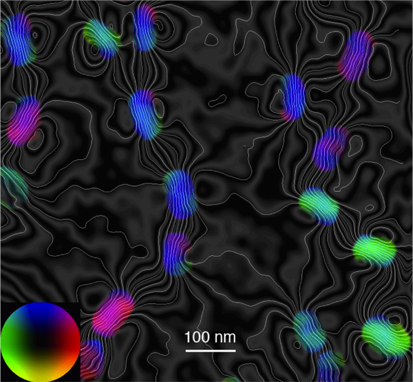
addition, advanced TEM techniques such as off-axis electron holography
and Z-contrast electron tomography are also used, in collaboration with
the group of Prof. Rafal Dunin-Borkowski. Using electron holography, it
is possible to both qualitatively and quantitatively characterize the
magnetic properties (such as magnetic induction mapping, measurement of
magnetic moments, coercive fields, etc.). |
 |
| |
|
|
Currently involved in this research at the UP: Éva Bereczk-Tompa, PhD student; Dr. Ilona Nyirő-Kósa, senior scientist. |
| |
|
|
Collaborations: Prof.
Rafal Dunin-Borkowski and Dr. András Kovács, Ernst Ruska-Centre for
Imaging and Spectroscopy with Electrons, Jülich Institute for Science
(off-axis electron holography for the characterization of magnetic
microstructures);
Dr. Damien Faivre, Max Planck Institute of Colloids and Surfaces
(syntheitic magnetic nanoparticles; Dr. Aleksander Rečnik, Jozef Stefan
Institute, Ljubljana (TEM of defects);
|
| |
|
|
Recent papers:
|
|
Taukulis, R., Widdrat, M., Kumari, M., Heinke, D., Rumpler, M., Tompa, É., Uebe, R., Kraupner, A., Cebers, A., Schüler, D., Pósfai, M., Hirt, A.M. and Faivre, D. (2015) Biological vs. synthetic and small vs. large magnetite nanoparticles as MRI contrast agents – a comprehensive physical and theoretical study. Magnetohydrodynamics, 51,721-747.
|
|
Kumari, M., Hirt, A.M., Uebe, R., Schüler, D., Tompa, É., Pósfai, M., Lorenz, W., Ahrentorp, F., Jonasson, C. and Johansson, C. (2015) Experimental mixtures of superparamagnetic and single-domain magnetite with respect to Day-Dunlop plots. Geochemistry, Geophysics, Geosystems, 16, 1739–1752, doi:10.1002/2015GC005744. |
|
Kumari, M., Widdrat, M., Tompa, É., Uebe, R., Schüler, D., Pósfai, M., Faivre, D. And Hirt, A.M. (2014) Distinguishing magnetic particle size of iron oxide nanoparticles with first-order reversal curves. Journal of Applied Physics, 116, 124304. |
|
Widdrat, M., Kumari, M., Tompa, É., Pósfai, M., Hirt, A. and Faivre, D. (2014)
Keeping nanoparticles fully functional: long term storage and alteration of magnetite. ChemPlusChem,
79, 1225-1233. doi: 10.1002/cplu.201402032. |
|
Rečnik, A., Nyirő-Kósa, I., Dódony, I. and Pósfai, M. (2013) Growth defects and epitaxy in Fe3O4 and γ-Fe2O3 nanocrystals. CrystEngComm, 15, 7539-7547. DOI: 10.1039/c3ce40873f. |
|
Pósfai, M., Kasama, T. and Dunin-Borkowski, R. (2013) Biominerals at the nanoscale: transmission electron microscopy methods for studying the special properties of biominerals.
In Minerals at the Nanoscale. European Mineralogical Union Notes in
Mineralogy, Vol. 14, F. Nieto and K. Livi eds., European Mineralogical
Union and the Mineralogical Society of Great Britain & Ireland,
London, 375-433. |
| |
|
| |
|
|
Synthesis of magnetic nanostructures |
| |
|
|
Magnetic structures with special shapes, particularly with elongated
morphologies, can have interesting applications because of their strong
magnetic anisotropy. While the formation of such structures is known in
nature (magnetotactic bacteria produce chains of magnetosomes), their
laboratory synthesis is difficult and requires high temperatures and/or
pressures. We aim at producing magnetic filaments using biomimetic
synthesis, by combining two known bacterial functions: magnetic
nanoparticle synthesis and filament formation. | |
| |
|
|
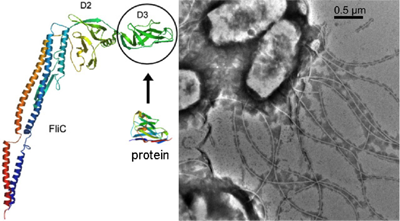
Many bacteria have flagella that are used for propelling the cell in an
aquatic environment. A flagellum is made of the flagellin protein that
contains a central, highly variable domain that can be replaced without
compromising the protein's stability. We aim at building filaments made
of mutagenized flagellin that contains iron-binding sites on its
surface. The iron-binding motifs are "imported" from the magnetosome
proteins of magnetotactic bacteria. The mutagenized filaments are then
used as templates for the precipitation of magnetite from solution.
This research is done in collaboration with the bio-nanotechnology
research group of Prof. Ferenc Vonderviszt. |
 |
| |
|
|
Currently involved in this research at the UP: Éva Bereczk-Tompa, PhD student;
Dr. Ferenc Vonderviszt, professor. |
| |
|
|
Collaborations: Dr. Damien Faivre and Victoria Reichel, Max Planck Institute for Colloids and Surfaces, Potsdam, Germany |
| |
|
| |
|
|
Recent papers:
|
|
Bereczk-Tompa, É., Vonderviszt, F., Horváth, B., Szalai, I., Pósfai, M. (2017) Biotemplated synthesis of magnetic filaments. Nanoscale, 9, 15062–15069. doi:10.1039/C7NR04842D.
|
|
Klein, Á., Kovács, M., Muskotál, A., Jankovics, H., Tóth, B., Pósfai, M., Vonderviszt, F. (2018) Nanobody-displaying flagellar nanotubes. Scientific Reports, 8:3584, doi:10.1038/s41598-018-22085-3.
|
|
Reichel, V., Kovács, A., Kumari, M., Bereczk-Tompa, É., Schneck, E., Diehle, P., Pósfai, M., Hirt, A.M., Duchamp, M., Dunin-Borkowski, R.E. and Faivre, D. (2017) Single crystalline superstructured stable single domain magnetite nanoparticles. Scientific Reports, 7, 45484, doi:10.1038/srep45484.
|
|
Bereczk-Tompa, É., Pósfai, M., Tóth, B., Vonderviszt, F. (2016) Magnetite-binding flagellar filaments displaying the MamI loop motif. ChemBioChem, DOI: 10.1002/cbic.201600377.
|
| |
|
| |
|
|
Carbonate formation in Lake Balaton |
| |
|
|
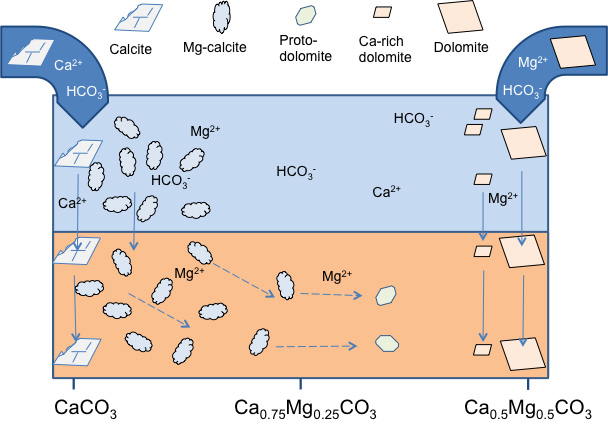
Lake Balaton has great economical and cultural importance in Hungary.
It is a large (70 km long) but shallow (on average 3.5 m deep),
calcareous lake with a sediment dominated by silt-size carbonate
grains. While most of the sediment consists of Mg-calcite that directly
precipitated from the water, calcite and dolomite from the erosion of the surrounding Triassic
carbonate hills also occurs in the sediment. In addition, "protodolomite" was also identified in the
sediment, probably forming diagenetically. | |
| |
|
|
Our research focuses on the carbonate mineralogy of the water and
sediment. We collect the freshly precipitated material using sediment
traps and also filter the suspended matter. Sediment cores were also
collected. We wish to determine the relationships between carbonate
composition and lakewater chemistry (primarily the Mg/Ca content), and
the origins of various carbonate minerals, including Mg-calcite,
micrometer-sized dolomite crystals and "protodolomite". It is also of
great interest wether planktonic organisms play any role in the
nucleation of the precipitating Mg-calcite grains, and to understand how the filtering
organisms (zooplankton and mussels) reprocess the primary carbonate
material. |
 |
| |
|
|
Currently involved in this research at the UP:
Dr. Ágnes Rostási, senior scientist; Dr. Ilona Nyirő-Kósa, senior scientist;
Melinda Fodor, PhD student; Zsombor Molnár, MSc student; Kornél Rácz, BSc student. |
| |
|
|
Collaborations: Prof. János Haas (dolomite formation), Eötvös L. University; Boglárka Topa and Dr. Tamás Weiszburg (XRD), Eötvös University. |
| |
|
|
Recent papers:
|
|
Nyirő-Kósa, I., Rostási, Á., Bereczk-Tompa, É., Cora, I., Koblar, M., Kovács, A., Pósfai, M. (2018) Nucleation and growth of Mg-bearing calcite in a shallow, calcareous lake. Earth and Planetary Science Letters, 496, 20-28. https://doi.org/10.1016/j.epsl.2018.05.029
|
|
Pósfai M., Rostási Á., Topa B., Molnár Zs., Nyirő-Kósa I., Bereczk-Tompa É., Fodor M., Cora I., Kovács A., Váczi T., Weiszburg T., Haas J. (2017) Karbonátásványok kiválása a Balatonban. (Precipitation of carbonate minerals in Lake Balaton – in Hungarian) 8. Kőzettani és Geokémiai Vándorgyűlés (Szihalom, szeptember 7-9.) Kiadványa, 143-146.
|
|
Pósfai, M., Nyirő-Kósa, I., Rostási, Á., Bereczk-Tompa, É., Cora, I., Koblar, M., Kovács, A. (2016) Nucleation, morphology, composition and structure of Mg-calcite, the dominant mineral in the mud of Lake Balaton Annual Meeting of the Hungarian Microscopy Society, Siófok, May 19-21.
|
|
Tompa, É., Nyirő-Kósa, I., Rostási, Á., Cserny, T. and Pósfai, M. (2014) Distribution and composition of Mg-calcite and dolomite in the water and sediments of Lake Balaton. Central European Geology,
57, 113-136. |
|
Nyirő-Kósa I., Tompa É., Rostási Á., G.-Tóth L., Nédli B.J., Balogh Cs., Cserny T. és Pósfai M. (2014) A Balaton karbonátásványai. Hidrológiai Közlöny, 94, 22-24. |
| |
|
| |
|
|
Atmospheric aerosol particles |
| |
|
|
Solid and liquid particles are ubiquitous constituents of Earth's
atmosphere. The particles have important roles in atmospheric
processes, since they serve as cloud condensation nuclei and scatter
and absorb both incoming shortwave and terrestrial infrared radiation.
In addition, the particles have strong influence on our environment by
reducing visibility and affecting health. In order to understand the
effects of atmospheric particles on our climate and environment, their
physical and chemical properties need to be known. | |
| |
|
|
We use transmission electron microscopy techniques for studying the
sizes, morphologies, compositions, structures and hygroscopic
properties of individual atmospheric particles. Since the particles
occur as distinct entities in the atmosphere, they can be regarded as
individual reaction vessels. Therefore, even though the bulk
composition of the atmospheric aerosol can be obtained using
established analytical methods, we also need to understand the physics
and chemistry of individual particles. In the past we have analyzed
representative particles from a large number of environmental settings
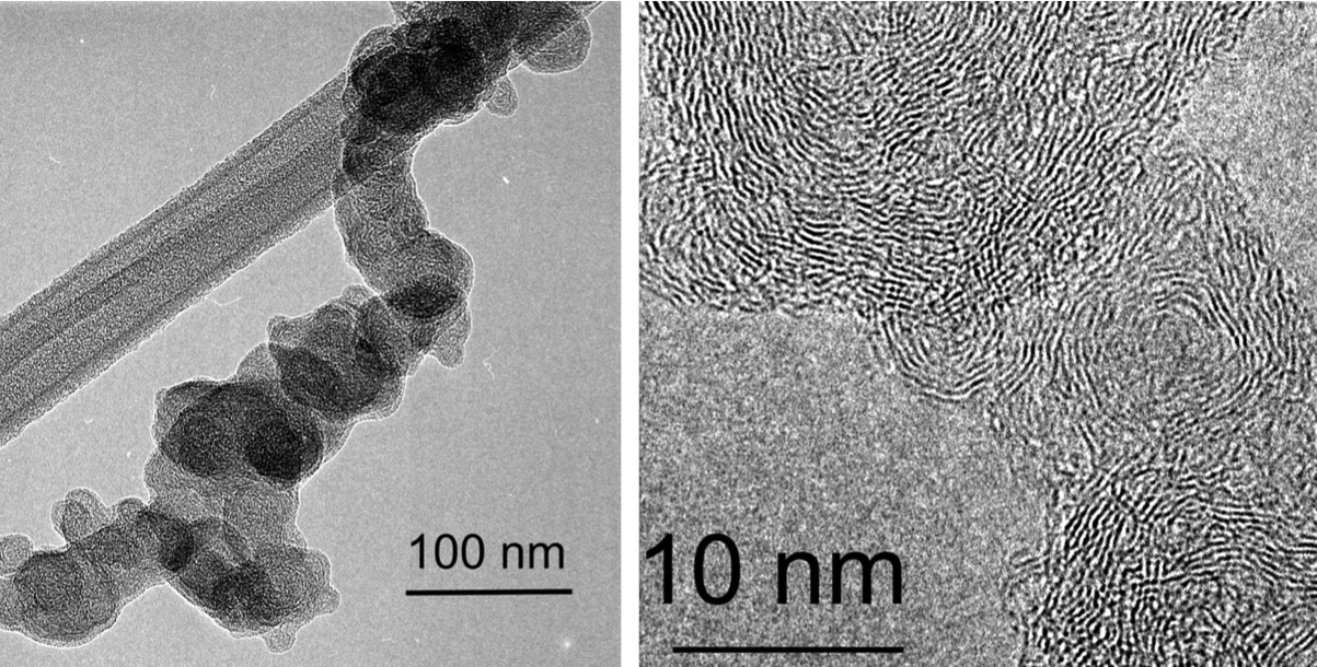
(remote oceanic, urban, rural). Currently, our main interest is in
understanding the physical properties and formation mechanisms of
particles produced by biomass burning. |
 |
| |
|
|
Currently involved in this research at the UP: Dr. Ilona Nyirő-Kósa, senior scientist. |
| |
|
|
Collaborations: Prof.
Peter Buseck, Arizona State University, Tempe, AZ;
Prof. András Gelencsér, Dr. Ágnes Molnár, Dr. András Hoffer and Ádám
Tóth, Air Chemistry Research Group of the Hungarian Academy of
Sciences, Veszprém. |
| |
|
|
Recent papers:
|
|
Hoffer, A., Tóth, Á., Pósfai, M., Chung, C.E., Gelencsér, A. (2017) Brown carbon absorption in the red and near-infrared spectral region. Atmospheric Measurement Techniques, 10, 2353-2359. https://doi.org/10.5194/amt-10-2353-2017. |
|
Hoffer, A., Tóth, Á., Nyirő-Kósa, I., Pósfai, M., Gelencsér, A. (2016) Light absorption properties of laboratory-generated tar ball particles. Atmospheric Chemistry and Physics, 16, 239-246, doi: 10.5194/acp-16-239-2016. |
|
Németh, Z., Pósfai, M., Nyirő-Kósa, I., Aalto, P., Kulmala, M. and Salma, I. (2015) Images and properties of individual nucleated particles. Atmospheric Environment, 123, 166-170. |
|
Buseck, P.R., Adachi, K., Gelencsér, A., Tompa, É. and Pósfai, M. (2014) Ns-soot: a material-based term for strongly light-absorbing carbonaceous particles. Aerosol Science and Technology, 48,
777-788. doi: 10.1080/02786826.2014.919374. |
|
Tóth, Á., Hoffer, A., Nyirő-Kósa, I., Pósfai, M. and Gelencsér, A. (2014) Atmospheric tar balls: aged primary droplets from biomass burning? Atmospheric Chemistry and Physics, 14, 6669-6675. doi:10.5194/acp-14-6669-2014. |
|
Pósfai, M. and Molnár, Á. (2013) Atmospheric aerosol particles: A mineralogical introduction.
In Environmental Mineralogy II. European Mineralogical Union Notes in
Mineralogy, Vol. 13, D. Vaughan and R. Wogelius eds., European
Mineralogical Union and the Mineralogical Society of Great Britain
& Ireland, London, 213-293. |
| |
|
| |
|
|